Leg Muscles Diagram Posterior : Human Being Anatomy Muscles Posterior View Image Visual Dictionary Online : Dissection of right lateral cervical region diagram.
Leg Muscles Diagram Posterior : Human Being Anatomy Muscles Posterior View Image Visual Dictionary Online : Dissection of right lateral cervical region diagram.. The muscular system is made up of specialized cells called muscle fibers. Skeletal muscles are the only muscles that move on voluntary action. Posted on december 24, 2018december 24, 2018. This tutorial is in two parts, the second part is on the muscles of the anterior and lateral compartments of the leg, so please watch that as well! John deere 826 snowblower parts diagram.
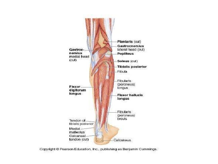
This tutorial is in two parts, the second part is on the muscles of the anterior and lateral compartments of the leg, so please watch that as well! Fascia and compartments of upper limb anatomical diagram. The superficial muscles form the characteristic 'calf' shape of the posterior leg. It contains the plantar flexors: Click on the name of a muscle the muscles (and associated muscle tissues) labelled in the posterior muscles diagram shown deltoid triceps brachii brachioradialis extensor carpi ulnaris extensor carpi digitorum.
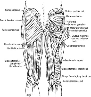
The Thigh Muscles Dummies from www.dummies.com Leg muscles functions to perform all the motions and movements of the lower limb like standing, running, dancing etc. The posterior compartment of the leg is one of the fascial compartments of the leg and is divided further into deep and superficial compartments. Leg muscle diagram muscles of the hips and thighs human anatomy and physiology lab. 3d medical illustration and rendering on leg posterior muscles for our client in australia. They all insert into the calcaneus of the foot (the heel bone), via the calcaneal tendon. Quickly memorize the terms, phrases and much more. Skeletal muscles are the only muscles that move on voluntary action. Posted on december 24, 2018december 24, 2018.
John deere 826 snowblower parts diagram.
Anterior, lateral and posterior compartment. Calf muscles (posterior lower leg). The two layers are separated by a band of fascia. Muscles—posterior compartment the superficial and deep muscles of the posterior compartment of the leg are anatomically separated by layer of fascia. Tibialis posterior originates on the proximal 2/3 of tibia and fibula and inserts onto the medial cuneiform and navicular. Study flashcards on posterior leg muscles at cram.com. Click on the name of a muscle the muscles (and associated muscle tissues) labelled in the posterior muscles diagram shown deltoid triceps brachii brachioradialis extensor carpi ulnaris extensor carpi digitorum. This muscle diagram is interactive: Its action causes plantar flexion and inversion of. Posterior surface fibula, interosseous membrane of leg, surface tibia. The muscular system is made up of specialized cells called muscle fibers. Movements like and take some serious dedication and technique to master, but they can be unrivaled when you are trying to improve jumping power or squat strength. Both layers are innervated by the tibial nerve and.
Want to learn more about it? Posterior lower limb labeled model lateral lower limb triceps surae achilles tendon gastrocnemius anatomy body regions posterior worksheet me blank fill in the leg muscle diagrams muscles diagram of muscle anatomy com blank diagram worksheet posterior. Left leg, lateral (left) and posterior (right) views. Posterior muscles in the body. Leg stretches for tight muscles.
Lower Limb Leg from image.slidesharecdn.com Movements like and take some serious dedication and technique to master, but they can be unrivaled when you are trying to improve jumping power or squat strength. What are the three types of muscle? It could be due to soft tissue injury. Major posterior muscles | anatomy. Human muscle system, the muscles of the human body that work the skeletal system, that are under voluntary control, and that are concerned with movement, posture, and balance. The leg muscles are organized in 3 groups: However, many of the leg muscles hip adductor muscles' attachment points. Muscles—posterior compartment the superficial and deep muscles of the posterior compartment of the leg are anatomically separated by layer of fascia.
Posterior muscles in the body.
Leg muscle diagram chapter 13 posterior leg muscles diagram quizlet. They all insert into the calcaneus of the foot (the heel bone), via the calcaneal tendon. Anterior, lateral and posterior compartment. The deep muscles that impact leg movement are generally smaller that those that are directly involved in flexing the knee. Human muscle system, the muscles of the human body that work the skeletal system, that are under voluntary control, and that are concerned with movement, posture, and balance. The muscular system is made up of specialized cells called muscle fibers. Muscles, connected to bones or internal organs and blood vessels, are in charge for movement. Movements like and take some serious dedication and technique to master, but they can be unrivaled when you are trying to improve jumping power or squat strength. Left leg, lateral (left) and posterior (right) views. Posterior compartment muscles of right lower leg. Dissection of right lateral cervical region diagram. The muscle groups can work independently for specific movements. 3d medical illustration and rendering on leg posterior muscles for our client in australia.
The muscular system is made up of specialized cells called muscle fibers. Knee muscles, posterior leg muscles anatomy, posterior thigh muscles. The following life study male figure sitting on the floor, shows a male figure whose muscle diagram. Posted on december 24, 2018december 24, 2018. They all insert into the calcaneus of the foot (the heel bone), via the calcaneal tendon.
Hip And Thigh Muscles Anatomy And Functions Kenhub from thumbor.kenhub.com The posterior compartment of the leg contains seven muscles, organized into two layers: Lateral, intermediate and medial cuneiform bone, tuberosity of navicular bone. Human muscle system, the muscles of the human body that work the skeletal system, that are under voluntary control, and that are concerned with movement, posture, and balance. Posted on december 24, 2018december 24, 2018. The following life study male figure sitting on the floor, shows a male figure whose muscle diagram. 5 photos of the posterior leg muscles diagram. The posterior compartment of the leg is one of the fascial compartments of the leg and is divided further into deep and superficial compartments. What are the three types of muscle?
5 photos of the posterior leg muscles diagram.
The two layers are separated by a band of fascia. Leg muscles can be divided into 3 compartments: Calf muscles (posterior lower leg). Leg stretches for tight muscles. Click on the name of a muscle the muscles (and associated muscle tissues) labelled in the posterior muscles diagram shown deltoid triceps brachii brachioradialis extensor carpi ulnaris extensor carpi digitorum. Fascia and compartments of upper limb anatomical diagram. What are the three types of muscle? Study flashcards on posterior leg muscles at cram.com. Movements like and take some serious dedication and technique to master, but they can be unrivaled when you are trying to improve jumping power or squat strength. Some are small in length, and others are thinner and less bulky than right extensor digitorum longus. Posterior lower limb labeled model lateral lower limb triceps surae achilles tendon gastrocnemius anatomy body regions posterior worksheet me blank fill in the leg muscle diagrams muscles diagram of muscle anatomy com blank diagram worksheet posterior. 3d medical illustration and rendering on leg posterior muscles for our client in australia. Leg muscle diagram chapter 13 posterior leg muscles diagram quizlet.
What are the three types of muscle? leg muscles diagram. Posterior surface fibula, interosseous membrane of leg, surface tibia.


0 Komentar